Pheochromocytoma: Everything You Need to Know
Pheochromocytoma is a rare tumor that forms in the adrenal glands. Often hidden for years, it can suddenly trigger severe symptoms like high blood pressure, rapid heartbeat, and sweating. Understanding its causes, signs, and treatment options is crucial for early detection and effective management.
Understanding Pheochromocytoma
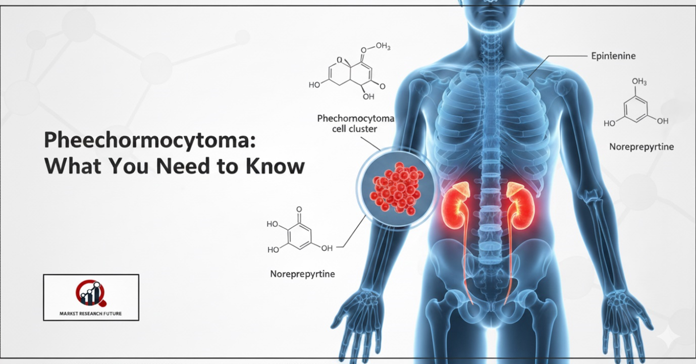
Pheochromocytoma is a rare tumor that develops in the adrenal glands, often lying undetected for years. When symptoms appear, they can be sudden and dramatic. This tumor causes the adrenal glands to produce excess adrenaline, also called the “fight-or-flight” hormone, affecting heart rate, blood pressure, and overall body function.
Recognizing the Symptoms
The signs of pheochromocytoma can be intense but sporadic. People may experience rapid heartbeat, high blood pressure, sweating, trembling, anxiety, or a sense of nervous tension. While memory and cognition usually remain normal, these symptoms can interfere with daily life. In some cases, the symptoms may mimic other conditions, including migraines, anxiety disorders, or even Parkinson’s disease.
Diagnosis and Testing
Diagnosing pheochromocytoma can be challenging because its symptoms overlap with many other conditions. Doctors typically begin with blood and urine tests to measure catecholamine levels. Imaging, such as CT or MRI scans of the abdomen and pelvis, helps locate the tumor and assess its size. Genetic testing may also be used to identify hereditary forms of the disease. Occasionally, the tumor is discovered incidentally during surgery for another condition.
Treatment and Management
Once diagnosed, treatment plans vary depending on the tumor’s size, location, and whether it is malignant. Surgery is the most common approach to remove the tumor. Patients may need to avoid strenuous activities, driving, or operating heavy machinery until treated, as sudden surges in adrenaline can cause dizziness or extreme anxiety. With proper medical care, most pheochromocytomas can be managed effectively.
Monitoring and Follow-Up
Regular monitoring is essential after diagnosis. Blood pressure, heart rate, and adrenal function are carefully tracked. Even when the tumor is benign, follow-up ensures any new growths are detected early. Malignant pheochromocytomas, though less common, require more extensive evaluation and treatment.
Conclusion
Pheochromocytoma is a rare but serious condition that affects the adrenal glands and can dramatically impact health if left untreated. Awareness of symptoms, timely diagnosis, and appropriate medical care are key. With proper management, patients can lead safe, active lives while minimizing the risks associated with this tumor.

Leave a Comment